Skin Cancer Removal & the Innovative Mohs Surgery

As board certified dermatologic and skin cancer surgeons, OC Skin Institute is the expert facility in Orange County for skin cancer removal and Mohs Surgery.  As we plan the best treatment for each of our patients, we consider the size and location of the skin cancer, the patient’s age and medical history, and the potential for scarring. Your overall health is our skin cancer surgeons’ primary concern, and their goal is to completely eliminate the skin cancer with as little recovery and scarring as possible.

What is Mohs Surgery?

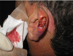
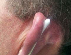
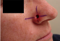
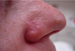
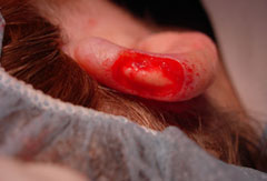
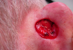
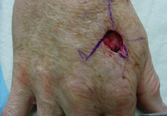
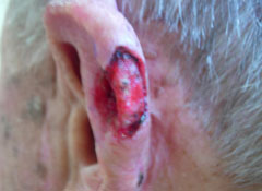
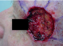
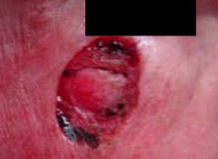
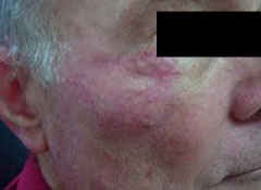
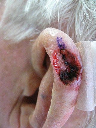
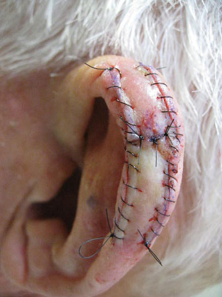
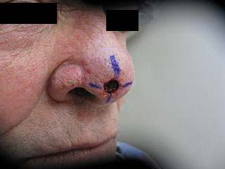
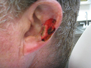
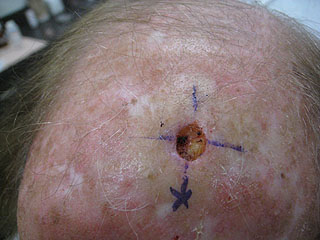
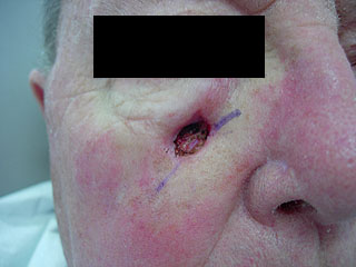
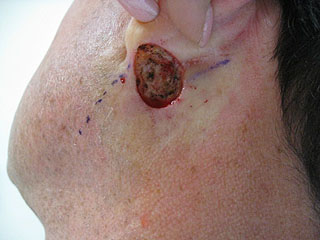
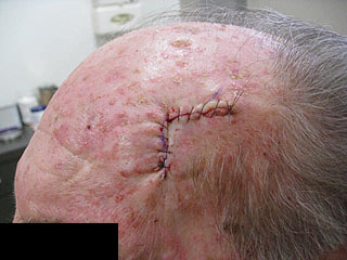
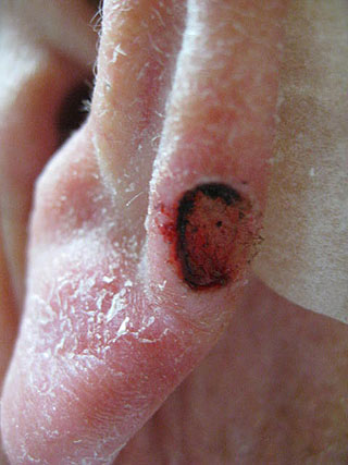
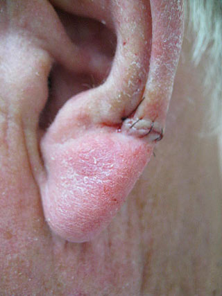
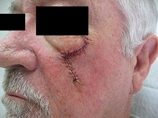
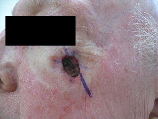
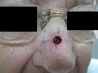
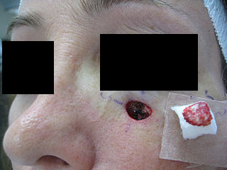
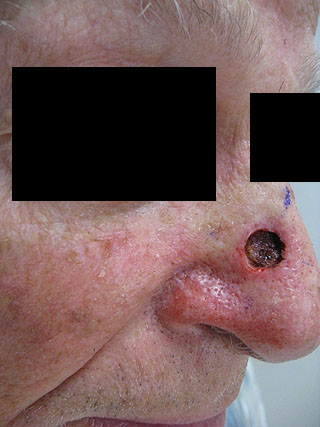
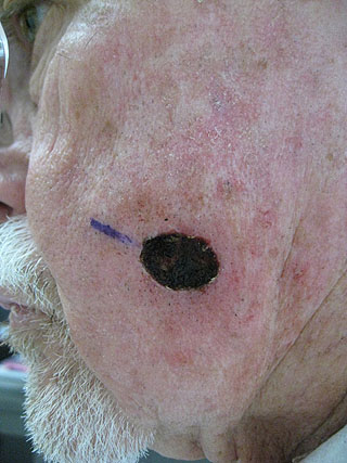
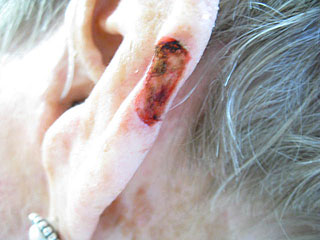
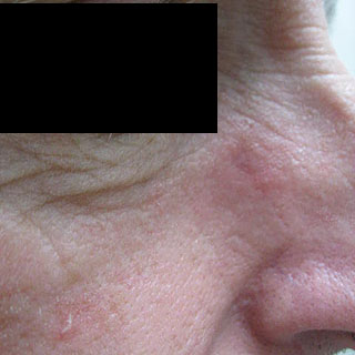
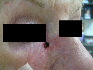
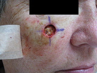
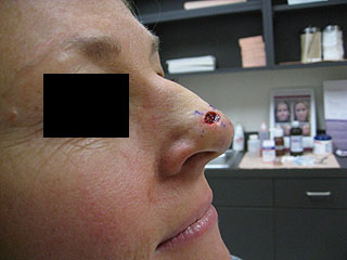
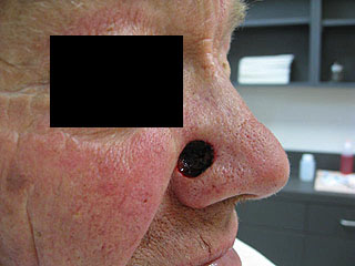
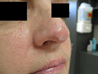
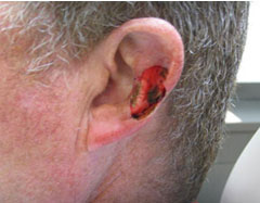
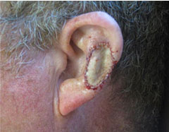
Mohs Surgery (named after its founder, Dr. Frederic Mohs), is an innovative micrographic technique for removing squamous cell and basal cell cancer. The cure rate using Mohs skin cancer surgery is very high, and recommended for large basal cell carcinomas on the body as well as for areas where recurrence of skin cancer is common such as on the scalp, face, hands, ears and nose. 

Some of the many advantages of Mohs surgery include:

  • Maximum preservation of healthy skin tissue
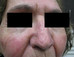
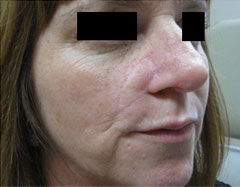
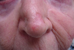
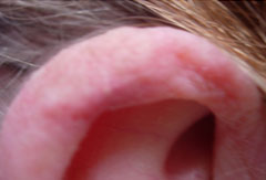
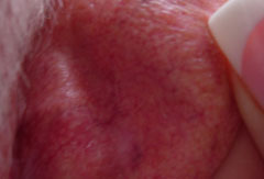
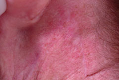
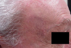
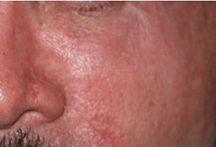
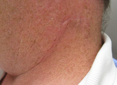
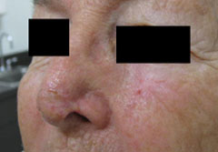
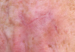
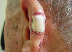
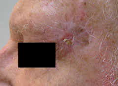
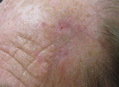
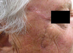
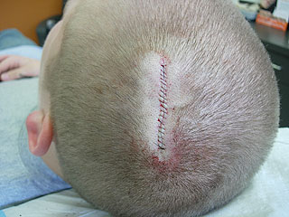
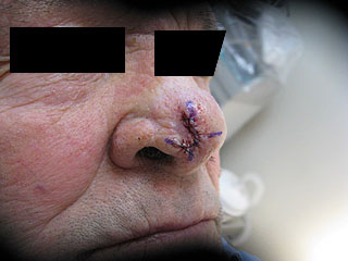
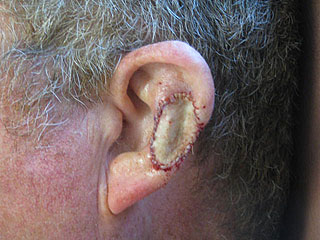
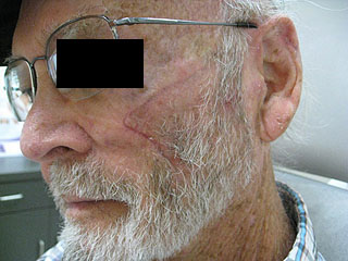
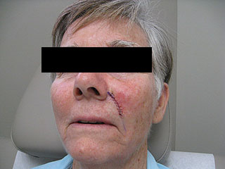
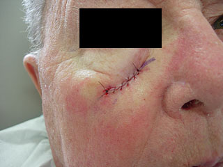
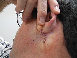
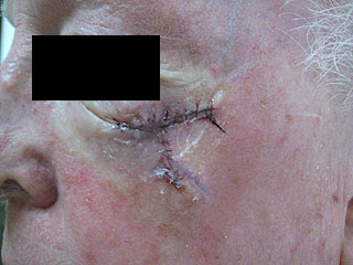
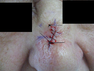
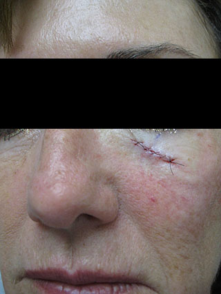
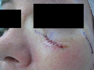
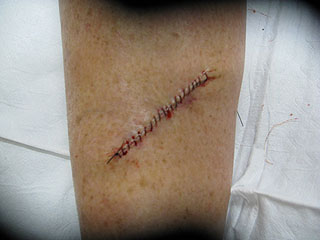
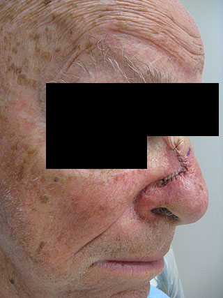
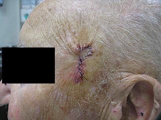
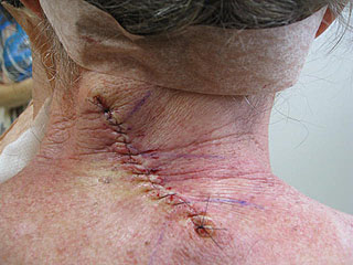
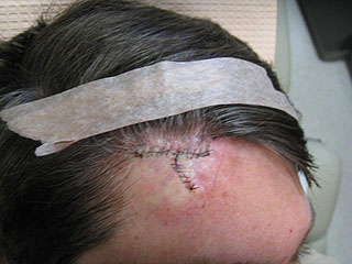
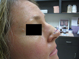
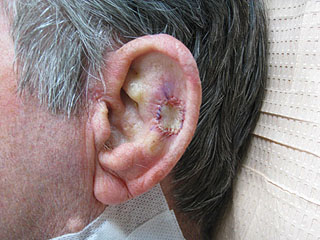
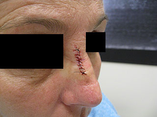
  • Minimal scarring
  • Its suitability for patients who have frequent skin cancers

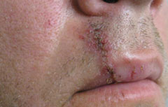
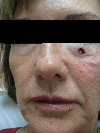
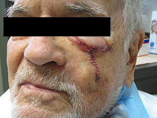
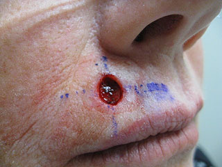
Older patients in poor health as well as patients with diabetes and other immune system deficiencies can be treated safely, as well as those needing skin cancer removal around the delicate areas of the eyes and mouth.

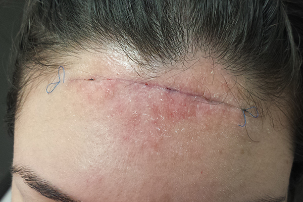
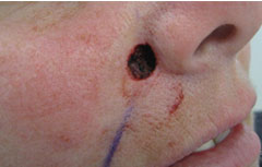
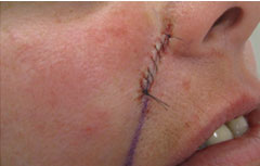
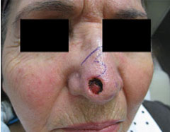
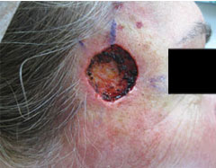
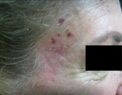
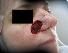
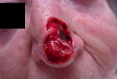
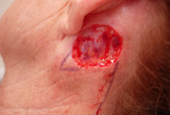
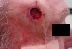
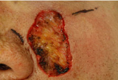
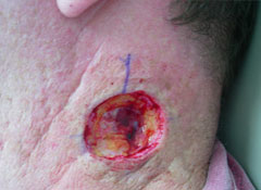
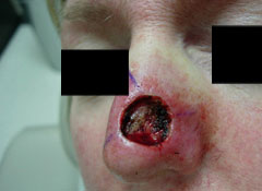
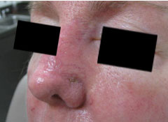
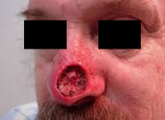
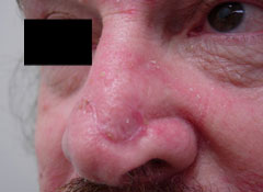
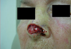
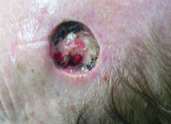
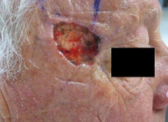
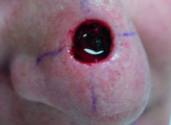
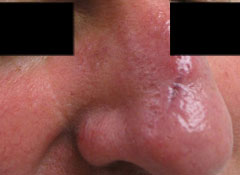
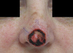
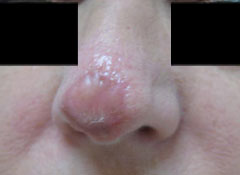
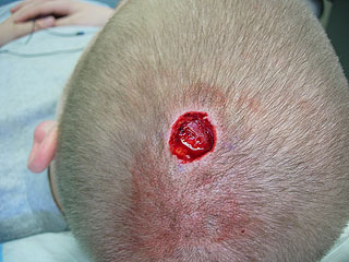
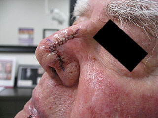
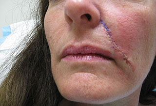
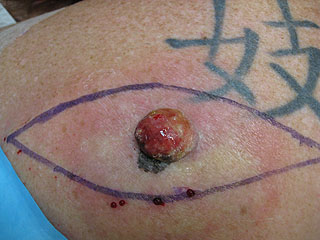
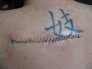
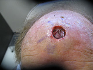
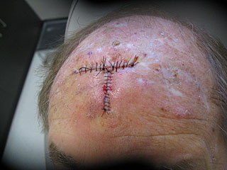
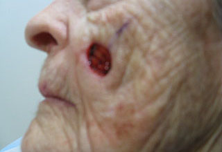
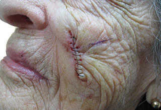
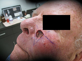
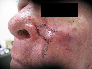
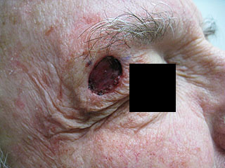
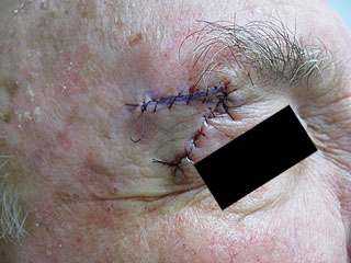
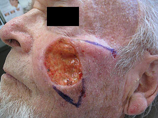
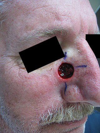
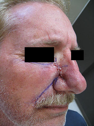
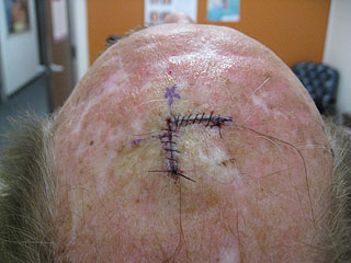
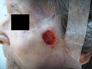
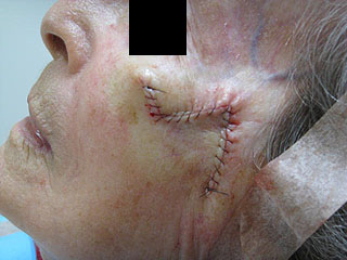
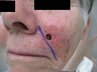
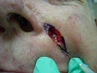
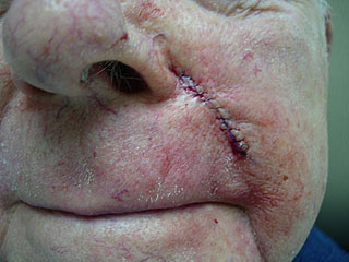
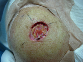
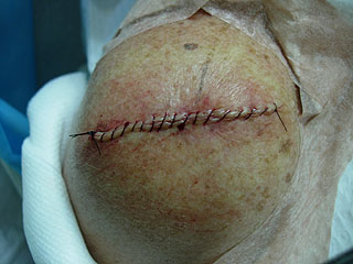
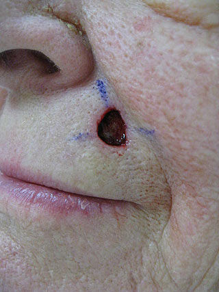
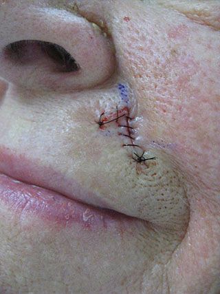
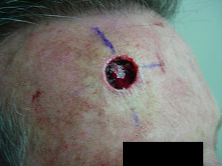
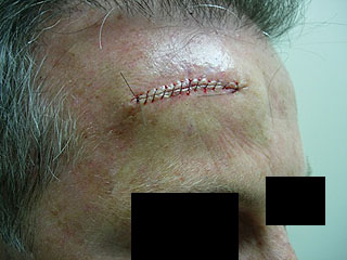
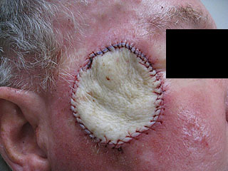
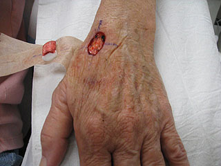
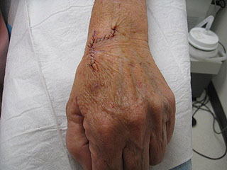
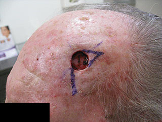
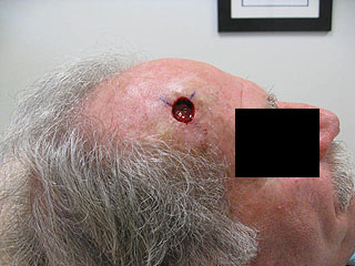
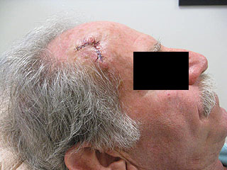
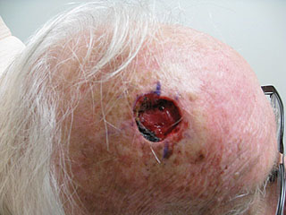
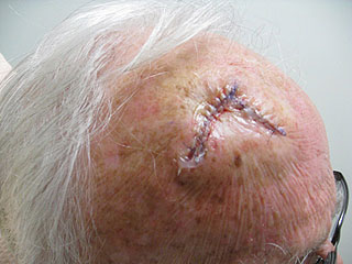
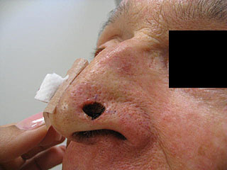
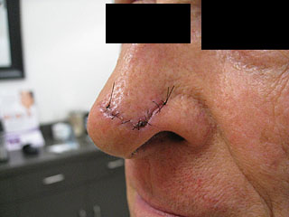
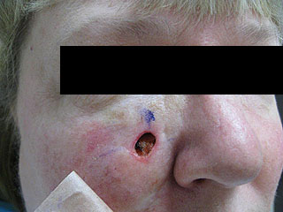
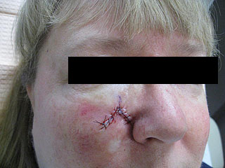
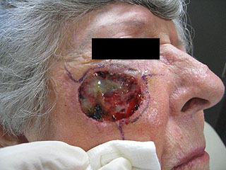
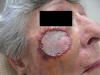
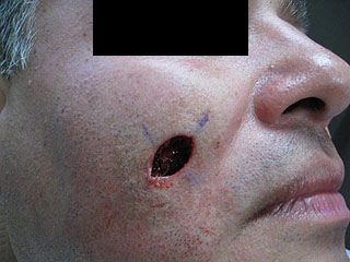
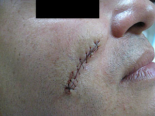
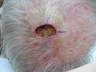
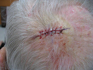
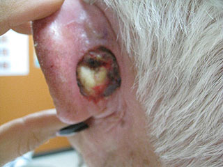
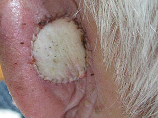
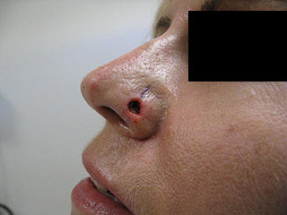
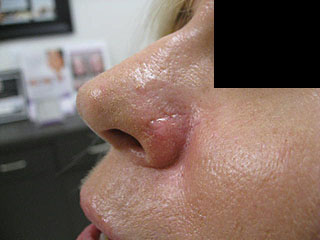
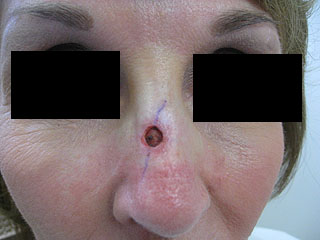
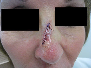
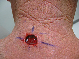
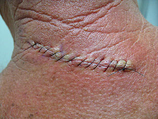
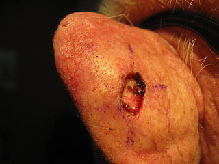
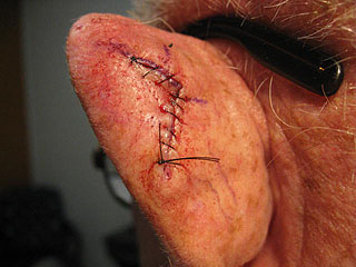
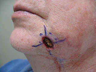
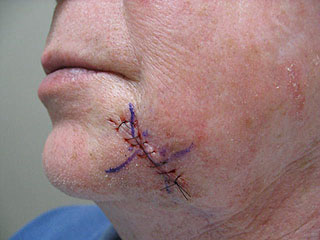
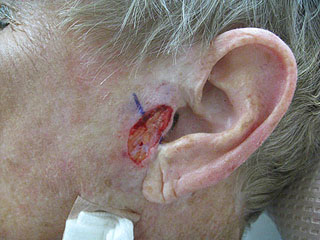
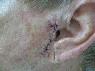
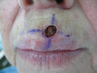
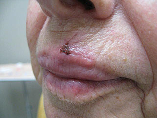
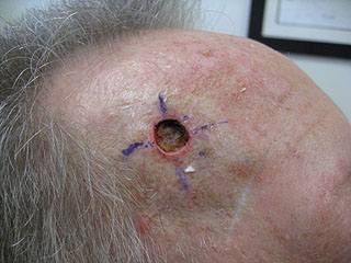
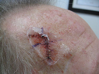
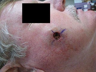
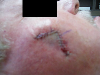
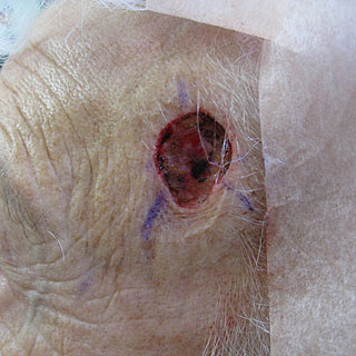
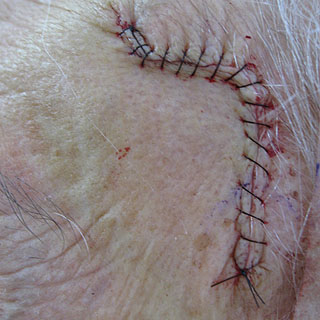
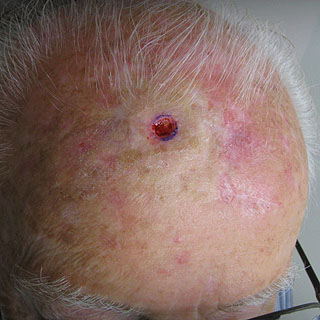
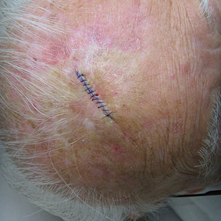
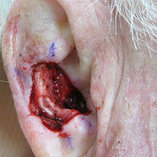
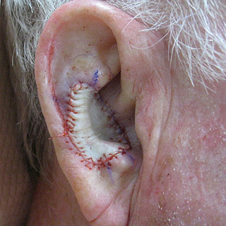
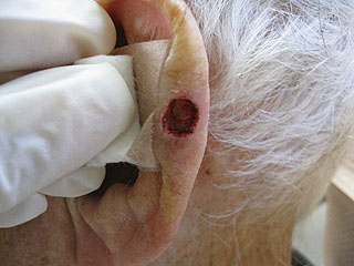
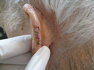
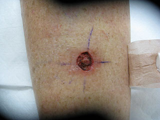
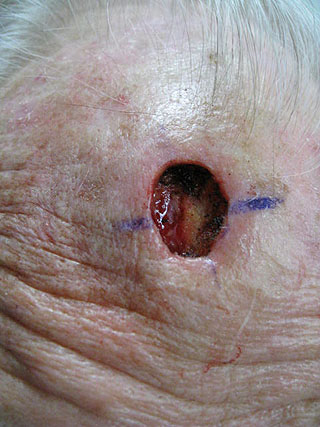
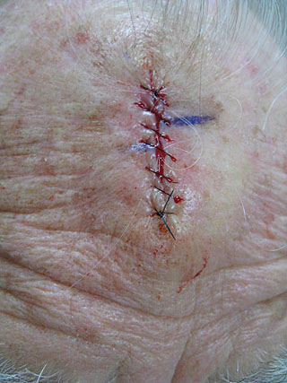
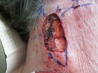
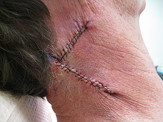
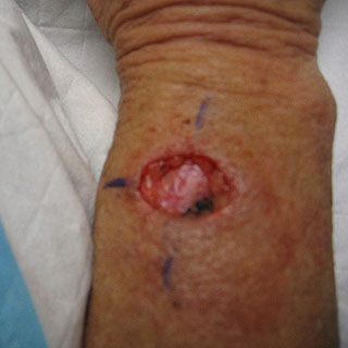
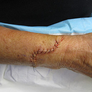
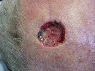
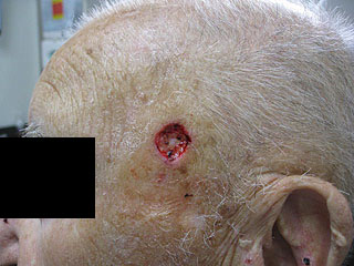
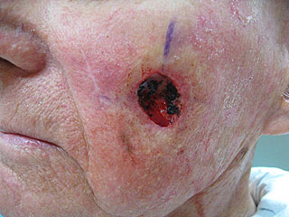
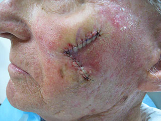
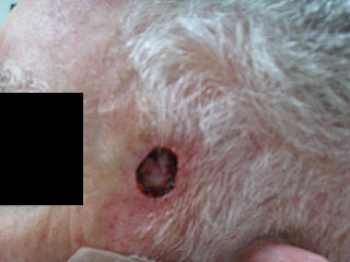
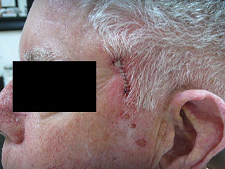
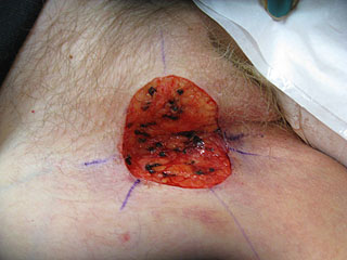
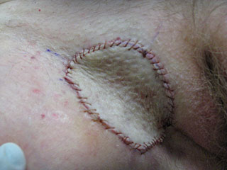
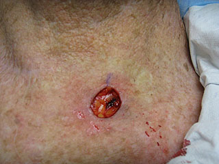
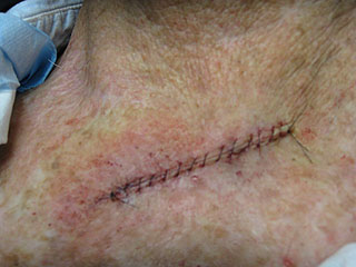
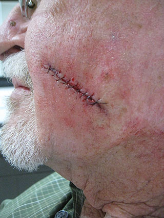
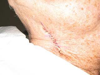
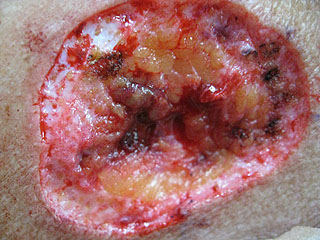
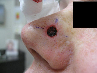
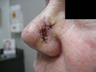
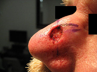
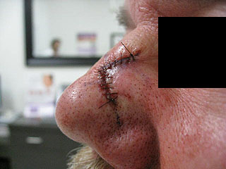
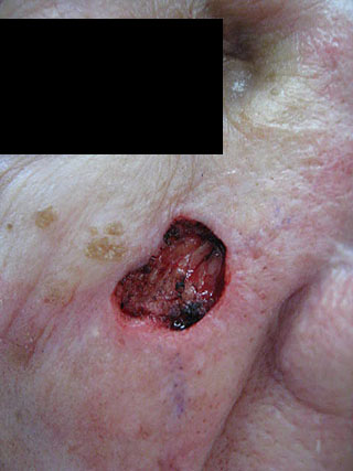
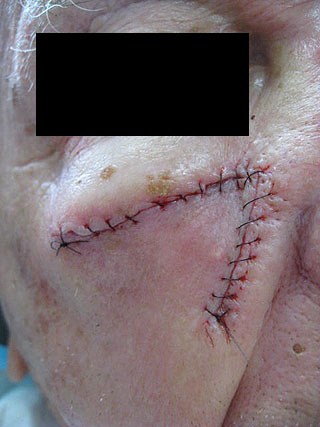
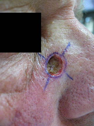
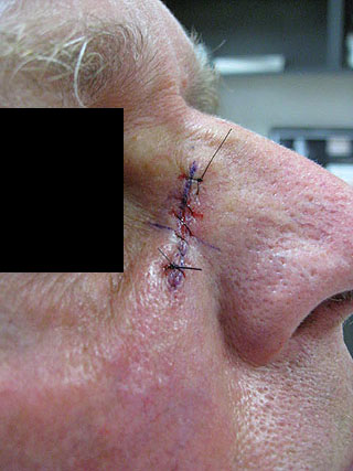
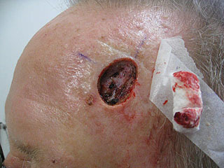
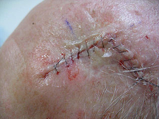
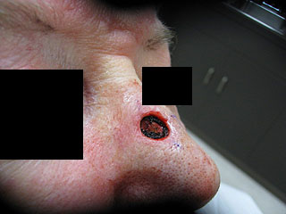
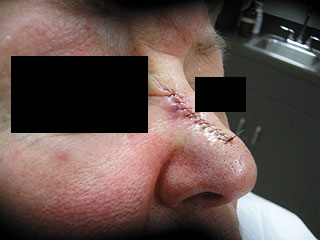
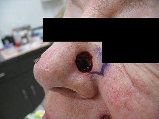
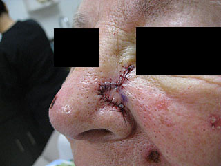
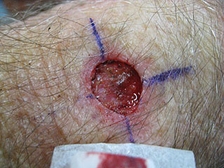
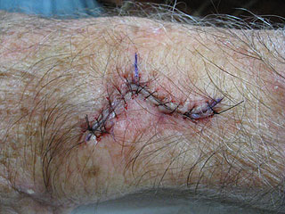
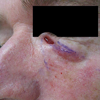
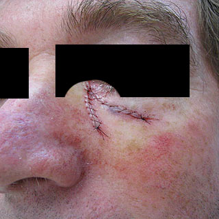
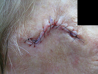
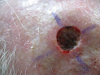
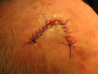
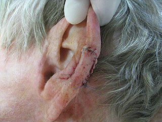
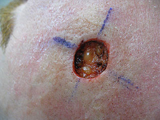
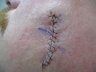
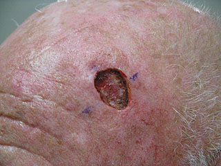
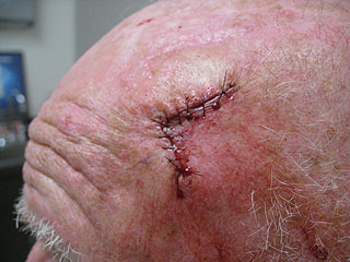
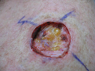
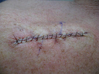
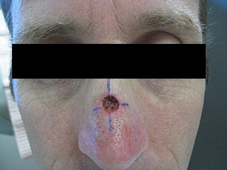
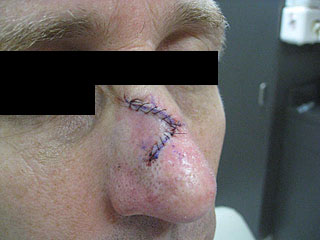
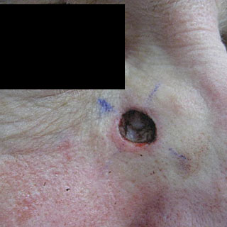
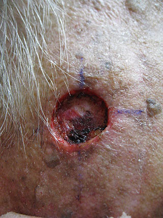
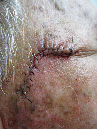
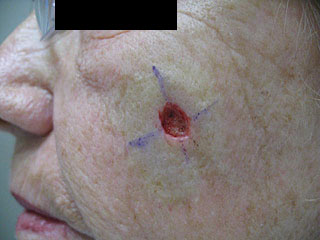
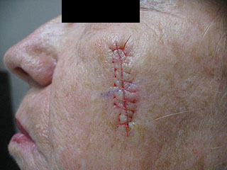
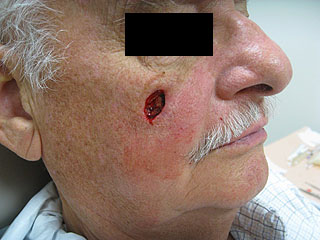
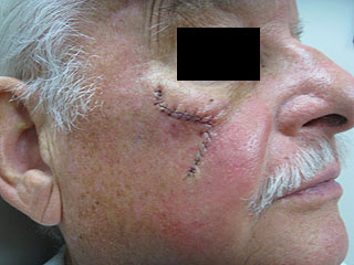
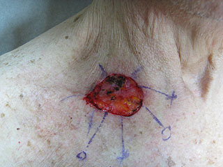
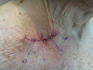
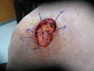
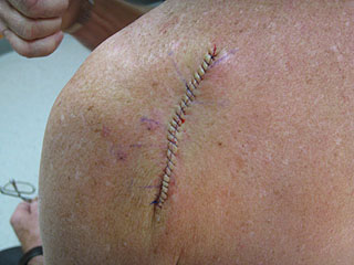
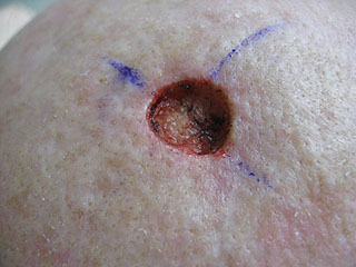
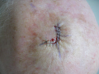
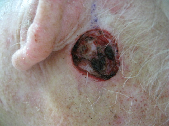
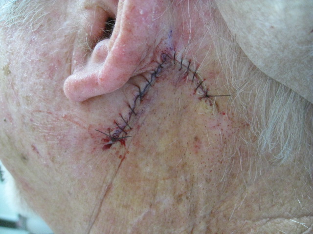
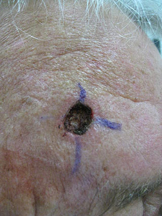
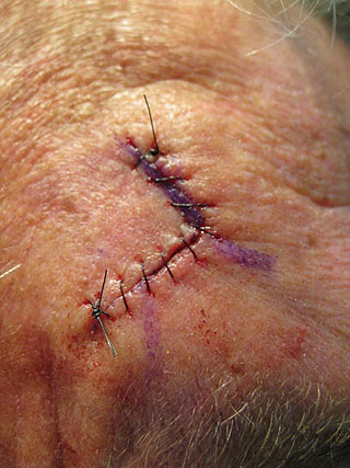
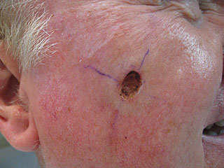
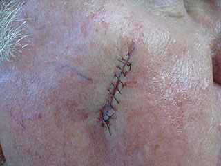
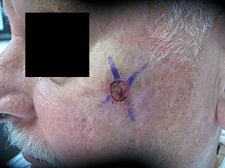
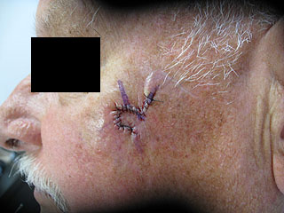
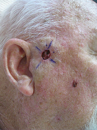
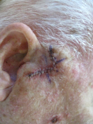
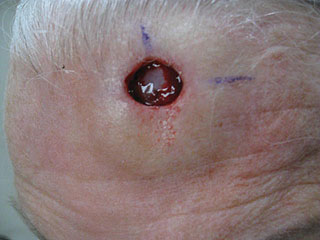
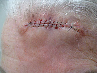
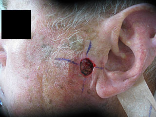
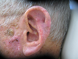
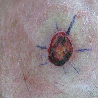
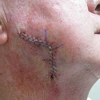
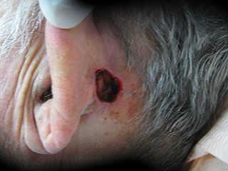
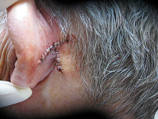
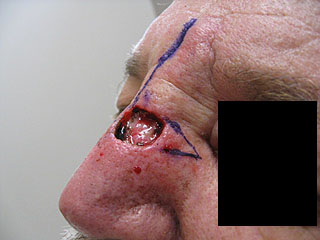
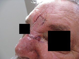
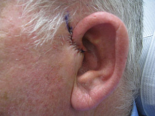
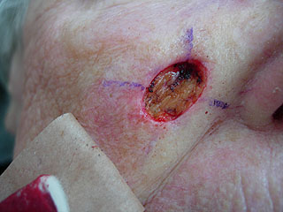
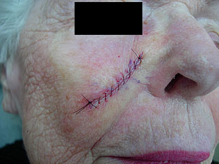
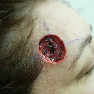
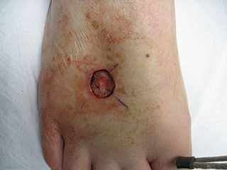
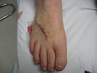
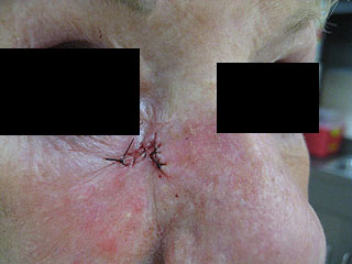
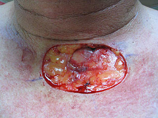
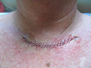
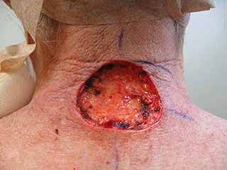
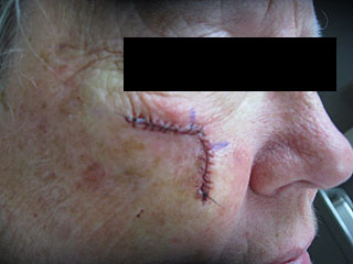
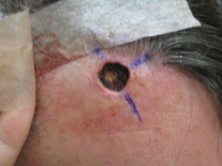
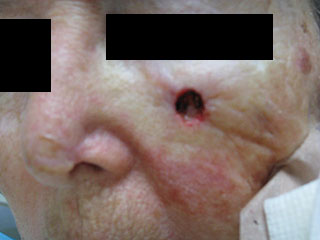
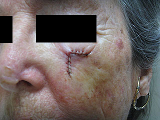
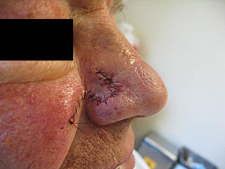
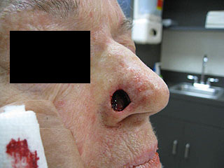
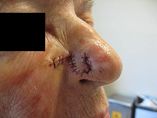
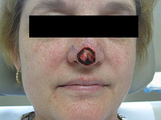
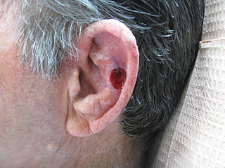
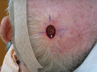
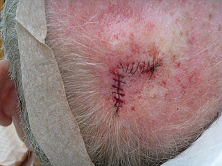
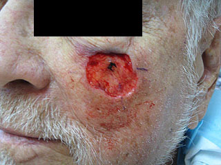
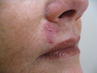
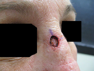
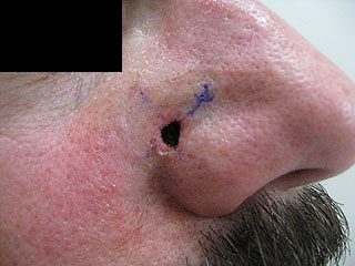
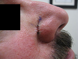
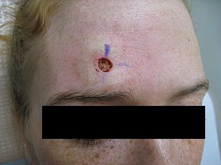
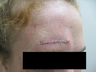
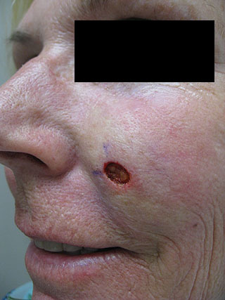
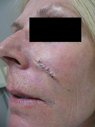
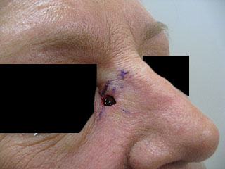
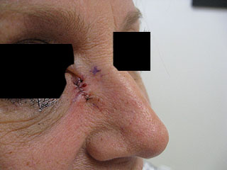
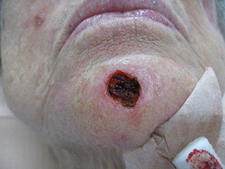
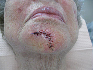
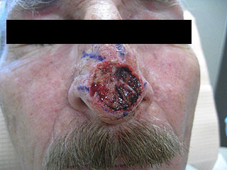
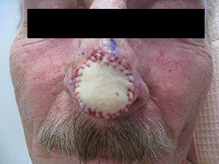
The procedure for skin cancer removal by Mohs surgery involves a microscopically controlled excision of small sections of the tumor, then immediately examining it under a microscope. If we identify cancer, we’ll remove more tissue until the section of skin cancer is clear and only normal skin is visible. The “hole” left by the basal cell cancer is then repaired the same or next day, leaving only a thin scar.

Other successful methods for skin cancer and precancerous treatment at OC Skin Institute include:

  • Cryosurgery: Typically, we apply liquid nitrogen to abnormal growths to freeze and kill the precancerous cells. This is best suited for precancerous lesions called actinic keratosis in order to prevent them from becoming full blown skin cancers.
  • Curettage and Desiccation: Best suited for small skin cancers on the trunk, arms or legs, this method involves the skin cancer surgeon using a spoon-like instrument called a curette to remove the abnormal cells. The surgeon then applies an electrical current to kill the remaining cancer cells and control bleeding. The tissue heals without the need for stitches.
  • Prescription Creams: Applied topically for several weeks, these formulations attack basal cells and stimulate the immune system. Although this method avoids surgery and can be applied by the patients themselves, it usually produces skin irritation and inflammation and discomfort. This method is reserved for patients who cannot undergo surgery or when a cancer or pre-cancer is observed to be very superficial and not invading deeper skin.
  • Radiation Therapy: For areas that are difficult to treat with skin cancer surgery, or in patients who cannot undergo surgery due to other health conditions, OC Skin Institute may refer a patient for radiation treatments for squamous or basal cell cancers.
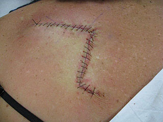
  • Surgical Removal: The skin cancer surgeon carefully removes the abnormal cells and stitches the area closed without immediate microscopic examination (as in Mohs skin cancer surgery). The specimen is sent to a lab to ensure that all of the margins are clear of skin cancer cells. This method is used for skin cancers on the body that are small and do not require Mohs Surgery.

At OC Skin Institute, we value our relationships with our patients and provide tailored expertise to each individual situation.  We have two locations throughout Orange County, including Santa Ana and Newport Beach, suited to provide full skin cancer treatment services for all patients. Please contact us for a consultation and qualified analysis of your skin cancer treatment needs.

Before & After Gallery

Real patients. Real results.
Case Study #1

Treatment: 32 year old female with 10 year history of tanning bed use. Basal carcinoma removed in 3 stages. Repaired with running subcuticular sutures for minimal scarring.

32 year old female with 10 year history of tanning bed use. Basal carcinoma removed in 3 stages. Repaired with running subcuticular sutures for minimal scarring. Before Photo Before
32 year old female with 10 year history of tanning bed use. Basal carcinoma removed in 3 stages. Repaired with running subcuticular sutures for minimal scarring. After Photo After
Case Study #2

Treatment: This is a case of a 42 year old gentleman with a large infiltrative basal cell carcinoma on the upper cutaneous lip extending onto the lip margin. Mohs surgery was complete in 2 stages followed by a lip wedge excision resulting in a thin linear scar.

This is a case of a 42 year old gentleman with a large infiltrative basal cell carcinoma on the upper cutaneous lip extending onto the lip margin. Mohs surgery was complete in 2 stages followed by a lip wedge excision resulting in a thin linear scar. Before Photo Before
This is a case of a 42 year old gentleman with a large infiltrative basal cell carcinoma on the upper cutaneous lip extending onto the lip margin. Mohs surgery was complete in 2 stages followed by a lip wedge excision resulting in a thin linear scar. After Photo After
Case Study #3

Treatment: This is a case of 40 year women with a basal cell carcinoma present in the nasal crease. Mohs surgery was complete in 1 level followed by reconstruction along the nasal labial fold. In this case, care was taken to orient the scar in the direction of the “smile line”.

This is a case of 40 year women with a basal cell carcinoma present in the nasal crease. Mohs surgery was complete in 1 level followed by reconstruction along the nasal labial fold. In this case, care was taken to orient the scar in the direction of the “smile line”. Before Photo Before
This is a case of 40 year women with a basal cell carcinoma present in the nasal crease. Mohs surgery was complete in 1 level followed by reconstruction along the nasal labial fold. In this case, care was taken to orient the scar in the direction of the “smile line”. After Photo After
Case Study #4

Treatment: This is a case of a 68 year old female with basal cell carcinoma on the nose. Mohs surgery was completed in 2 stages followed by a bilobed flap for reconstruction.

This is a case of a 68 year old female with basal cell carcinoma on the nose. Mohs surgery was completed in 2 stages followed by a bilobed flap for reconstruction. Before Photo Before
This is a case of a 68 year old female with basal cell carcinoma on the nose. Mohs surgery was completed in 2 stages followed by a bilobed flap for reconstruction. After Photo After
Case Study #5

Treatment: This is a case of a 70 year old gentleman with a squamous cell carcinoma on the left ear. Mohs surgery was performed and completed in 2 stages followed by an H-plasty helical rim repair.

This is a case of a 70 year old gentleman with a squamous cell carcinoma on the left ear. Mohs surgery was performed and completed in 2 stages followed by an H-plasty helical rim repair. Before Photo Before
This is a case of a 70 year old gentleman with a squamous cell carcinoma on the left ear. Mohs surgery was performed and completed in 2 stages followed by an H-plasty helical rim repair. After Photo After
Case Study #6

Treatment: This is a case of a 65 year old female with a large infiltrative basal cell carcinoma on the temple. Mohs surgery was completed in 4 levels followed by a rhombic flap for reconstruction. The final outcome resulted in a barely visible scar.

This is a case of a 65 year old female with a large infiltrative basal cell carcinoma on the temple. Mohs surgery was completed in 4 levels followed by a rhombic flap for reconstruction. The final outcome resulted in a barely visible scar. Before Photo Before
This is a case of a 65 year old female with a large infiltrative basal cell carcinoma on the temple. Mohs surgery was completed in 4 levels followed by a rhombic flap for reconstruction. The final outcome resulted in a barely visible scar. After Photo After
Case Study #7

Treatment: This is a case of 38 year old young women with a large, infiltrative basal cell carcinoma on the right cheek extending medially onto the nasal sidewall and ala. Mohs surgery was completed in 5 stages followed by an island pedicle flap. Care was taken to hide the scar within the adjacent “smile line”

This is a case of 38 year old young women with a large, infiltrative basal cell carcinoma on the right cheek extending medially onto the nasal sidewall and ala. Mohs surgery was completed in 5 stages followed by an island pedicle flap. Care was taken to hide the scar within the adjacent “smile line” Before Photo Before
This is a case of 38 year old young women with a large, infiltrative basal cell carcinoma on the right cheek extending medially onto the nasal sidewall and ala. Mohs surgery was completed in 5 stages followed by an island pedicle flap. Care was taken to hide the scar within the adjacent “smile line” After Photo After
Case Study #8

Treatment: This is a case of a small alar basal cell carcinoma on a young female patient concerned with the cosmetic outcome. A single stage of Mohs cleared the lesion followed by a small single advancement flap. Care was taken to avoid alar and nasal tip distortion.

This is a case of a small alar basal cell carcinoma on a young female patient concerned with the cosmetic outcome. A single stage of Mohs cleared the lesion followed by a small single advancement flap. Care was taken to avoid alar and nasal tip distortion. Before Photo Before
This is a case of a small alar basal cell carcinoma on a young female patient concerned with the cosmetic outcome. A single stage of Mohs cleared the lesion followed by a small single advancement flap. Care was taken to avoid alar and nasal tip distortion. After Photo After
Case Study #9

Treatment: This is a basal cell carcinoma defect after 6 levels of Mohs surgery. The defect was repair with a full thickness skin graft.

This is a basal cell carcinoma defect after 6 levels of Mohs surgery. The defect was repair with a full thickness skin graft. Before Photo Before
This is a basal cell carcinoma defect after 6 levels of Mohs surgery. The defect was repair with a full thickness skin graft. After Photo After
Case Study #10

Treatment: This is a basal cell carcinoma on the ear treated with 1 level of Mohs surgery followed by a full thickness skin graft.

This is a basal cell carcinoma on the ear treated with 1 level of Mohs surgery followed by a full thickness skin graft. Before Photo Before
This is a basal cell carcinoma on the ear treated with 1 level of Mohs surgery followed by a full thickness skin graft. After Photo After
Case Study #11

Treatment: This is a case of a 75 year old gentleman with basal cell carcinoma on the posterior ear treated with Mohs surgery in 2 levels and repaired using a rhombic flap.

This is a case of a 75 year old gentleman with basal cell carcinoma on the posterior ear treated with Mohs surgery in 2 levels and repaired using a rhombic flap. Before Photo Before
This is a case of a 75 year old gentleman with basal cell carcinoma on the posterior ear treated with Mohs surgery in 2 levels and repaired using a rhombic flap. After Photo After
Case Study #12

Treatment: This is a case of a 72 year old female with an infiltrative basal cell carcinoma present on the left post auricular region. Mohs surgery was employed and the tumor was cleared in 4 levels, followed by a large single advancement flap for reconstruction.

This is a case of a 72 year old female with an infiltrative basal cell carcinoma present on the left post auricular region. Mohs surgery was employed and the tumor was cleared in 4 levels, followed by a large single advancement flap for reconstruction. Before Photo Before
This is a case of a 72 year old female with an infiltrative basal cell carcinoma present on the left post auricular region. Mohs surgery was employed and the tumor was cleared in 4 levels, followed by a large single advancement flap for reconstruction. After Photo After
Case Study #13

Treatment: This is a case of basal cell carcinoma present on the right temple. Mohs surgery was employed followed by an O-T repair for reconstruction.

This is a case of basal cell carcinoma present on the right temple. Mohs surgery was employed followed by an O-T repair for reconstruction. Before Photo Before
This is a case of basal cell carcinoma present on the right temple. Mohs surgery was employed followed by an O-T repair for reconstruction. After Photo After
Case Study #14

Treatment: This is a case of a young gentleman with recurrent infiltrative basal cell carcinoma previous treated with Mohs surgery at another office. The patient underwent Mohs surgery with Dr. Nakhla and was cleared in 5 stages. The large defect was repaired using a single advancement flap carefully planned with all incisions placed along natural skin creases resulting in a nearly scarless surgery.

This is a case of a young gentleman with recurrent infiltrative basal cell carcinoma previous treated with Mohs surgery at another office. The patient underwent Mohs surgery with Dr. Nakhla and was cleared in 5 stages. The large defect was repaired using a single advancement flap carefully planned with all incisions placed along natural skin creases resulting in a nearly scarless surgery. Before Photo Before
This is a case of a young gentleman with recurrent infiltrative basal cell carcinoma previous treated with Mohs surgery at another office. The patient underwent Mohs surgery with Dr. Nakhla and was cleared in 5 stages. The large defect was repaired using a single advancement flap carefully planned with all incisions placed along natural skin creases resulting in a nearly scarless surgery. After Photo After
Case Study #15

Treatment: This is a case of a gentleman with a large basal cell carcinoma on the right neck requiring 4 levels of Mohs surgery followed by a large single advancement flap. Care was taken to place the incisions with a wrinkle on the neck resulting in minimally visible scarring.

This is a case of a gentleman with a large basal cell carcinoma on the right neck requiring 4 levels of Mohs surgery followed by a large single advancement flap.  Care was taken to place the incisions with a wrinkle on the neck resulting in minimally visible scarring. Before Photo Before
This is a case of a gentleman with a large basal cell carcinoma on the right neck requiring 4 levels of Mohs surgery followed by a large single advancement flap.  Care was taken to place the incisions with a wrinkle on the neck resulting in minimally visible scarring. After Photo After
Case Study #16

Treatment: This is a case of a young female patient with a large basal cell cancer on the nose. Mohs surgery was used to remove the tumor in 3 levels, followed by reconstruction using a hatchet flap resulting in an excellent cosmetic outcome considering the size and location of the tumor.

This is a case of a young female patient with a large basal cell cancer on the nose. Mohs surgery was used to remove the tumor in 3 levels, followed by reconstruction using a hatchet flap resulting in an excellent cosmetic outcome considering the size and location of the tumor. Before Photo Before
This is a case of a young female patient with a large basal cell cancer on the nose. Mohs surgery was used to remove the tumor in 3 levels, followed by reconstruction using a hatchet flap resulting in an excellent cosmetic outcome considering the size and location of the tumor. After Photo After
Case Study #17

Treatment: This is another case of a large tumor on the nose. This lesion was a squamous cell carcinoma of the keratoacanthoma type. Mohs surgery was employed followed by reconstruction using a tri-lobed flap from borrowed skin on the opposing side of the nose.

This is another case of a large tumor on the nose. This lesion was a squamous cell carcinoma of the keratoacanthoma type. Mohs surgery was employed followed by reconstruction using a tri-lobed flap from borrowed skin on the opposing side of the nose. Before Photo Before
This is another case of a large tumor on the nose. This lesion was a squamous cell carcinoma of the keratoacanthoma type. Mohs surgery was employed followed by reconstruction using a tri-lobed flap from borrowed skin on the opposing side of the nose. After Photo After
Case Study #18

Treatment: This is a basal cell carcinoma present on the nasal tip of a 83 year old female after 3 levels of Mohs surgery followed by a full thickness skin graft.

This is a basal cell carcinoma present on the nasal tip of a 83 year old female after 3 levels of Mohs surgery followed by a full thickness skin graft. Before Photo Before
This is a basal cell carcinoma present on the nasal tip of a 83 year old female after 3 levels of Mohs surgery followed by a full thickness skin graft. After Photo After
Case Study #19

Treatment: This is a case of a squamous cell carcinoma present on the dorsal surface of the hand after 1 level of Mohs surgery and a single advancement flap for repair.

This is a case of a squamous cell carcinoma present on the dorsal surface of the hand after 1 level of Mohs surgery and a single advancement flap for repair. Before Photo Before
This is a case of a squamous cell carcinoma present on the dorsal surface of the hand after 1 level of Mohs surgery and a single advancement flap for repair. After Photo After
Case Study #20

Treatment: Mohs surgery

Patient Details: This is a case of basal cell carcinoma following mohs surgery and a full thickness skin graft on the helical rim of the ear.

Mohs surgery Before Photo Before
Mohs surgery After Photo After
Case Study #21

Treatment: Basal cell carcinoma on the left temple of a 78 year old gentleman after 4 levels of Mohs micrographic surgery. The defect was allowed to contract and granulate for 2 weeks followed by repair using a full thickness skin graft.

Basal cell carcinoma on the left temple of a 78 year old gentleman after 4 levels of Mohs micrographic surgery. The defect was allowed to contract and granulate for 2 weeks followed by repair using a full thickness skin graft. Before Photo Before
Basal cell carcinoma on the left temple of a 78 year old gentleman after 4 levels of Mohs micrographic surgery. The defect was allowed to contract and granulate for 2 weeks followed by repair using a full thickness skin graft. After Photo After
Case Study #22

Treatment: Basal cell carcinoma on the left temple treated with 3 levels of Mohs micrographic surgery followed by repair using an O-L single advancement flap.

Basal cell carcinoma on the left temple treated with 3 levels of Mohs micrographic surgery followed by repair using an O-L single advancement flap. Before Photo Before
Basal cell carcinoma on the left temple treated with 3 levels of Mohs micrographic surgery followed by repair using an O-L single advancement flap. After Photo After
Case Study #23

Treatment: This is a case of basal cell carcinoma treated with Mohs micrographic surgery followed by reconstruction using an O-T bilateral advancement flap.

This is a case of basal cell carcinoma treated with Mohs micrographic surgery followed by reconstruction using an O-T bilateral advancement flap. Before Photo Before
This is a case of basal cell carcinoma treated with Mohs micrographic surgery followed by reconstruction using an O-T bilateral advancement flap. After Photo After
Case Study #24

Treatment: Squamous cell carcinoma on the nose after 1 stage of Mohs micrographic surgery followed by linear closure.

Squamous cell carcinoma on the nose after 1 stage of Mohs micrographic surgery followed by linear closure. Before Photo Before
Squamous cell carcinoma on the nose after 1 stage of Mohs micrographic surgery followed by linear closure. After Photo After
Case Study #25

Treatment: This is a case of infiltrative basal cell carcinoma on a young female patient’s nose. The tumor extended all the way to the cartilage after treatment with 5 levels of Mohs micrographic surgery followed by a full thickness skin graft.

This is a case of infiltrative basal cell carcinoma on a young female patient’s nose. The tumor extended all the way to the cartilage after treatment with 5 levels of Mohs micrographic surgery followed by a full thickness skin graft. Before Photo Before
This is a case of infiltrative basal cell carcinoma on a young female patient’s nose. The tumor extended all the way to the cartilage after treatment with 5 levels of Mohs micrographic surgery followed by a full thickness skin graft. After Photo After
Case Study #26

Treatment: This is a case of infiltrative basal cell carcinoma on the right infraorbital region after 5 levels of Mohs micrographic surgery. The defect was repaired used a large single advancement flap from the zygomatic area.

This is a case of infiltrative basal cell carcinoma on the right infraorbital region after 5 levels of Mohs micrographic surgery. The defect was repaired used a large single advancement flap from the zygomatic area. Before Photo Before
This is a case of infiltrative basal cell carcinoma on the right infraorbital region after 5 levels of Mohs micrographic surgery. The defect was repaired used a large single advancement flap from the zygomatic area. After Photo After
Case Study #27

Treatment: This is a case of basal cell carcinoma on the scalp of a young man after 2 levels of Mohs surgery followed by linear closure.

This is a case of basal cell carcinoma on the scalp of a young man after 2 levels of Mohs surgery followed by linear closure. Before Photo Before
This is a case of basal cell carcinoma on the scalp of a young man after 2 levels of Mohs surgery followed by linear closure. After Photo After
Case Study #28

Treatment: Squamous cell carcinoma on the nasal tip after Mohs surgery followed by a hatchet flap.

Squamous cell carcinoma on the nasal tip after Mohs surgery followed by a hatchet flap. Before Photo Before
Squamous cell carcinoma on the nasal tip after Mohs surgery followed by a hatchet flap. After Photo After
Case Study #29

Treatment: This is a case of sclerosing basal cell carcinoma present on the upper cutaneous lip after 3 levels of Mohs micrographic surgery. The defect was extended laterally onto the nasal labial fold (smile line) to hide the scar within the natural fold.

This is a case of sclerosing basal cell carcinoma present on the upper cutaneous lip after 3 levels of Mohs micrographic surgery. The defect was extended laterally onto the nasal labial fold (smile line) to hide the scar within the natural fold. Before Photo Before
This is a case of sclerosing basal cell carcinoma present on the upper cutaneous lip after 3 levels of Mohs micrographic surgery. The defect was extended laterally onto the nasal labial fold (smile line) to hide the scar within the natural fold. After Photo After
Case Study #30

Treatment: Squamous cell carcinoma on the ear repaired with a bilateral H-plasty helical rim flap.

Squamous cell carcinoma on the ear repaired with a bilateral H-plasty helical rim flap. Before Photo Before
Squamous cell carcinoma on the ear repaired with a bilateral H-plasty helical rim flap. After Photo After
Case Study #31

Treatment: Melanoma Excisional Biopsy

Patient Details: This is a patient that presented to OC Skin Institute with a large tumor on the back. The clinical appearance of the lesion warranted an excisional biopsy of the entire lesion with 1cm margins which was diagnosed as invasive melanoma.

Melanoma Excisional Biopsy Before Photo Before
Melanoma Excisional Biopsy After Photo After
Case Study #32

Treatment: This is a case of basal cell carcinoma on the scalp after 5 levels of Mohs surgery. The defect was closed with an O-T advancement flap.

This is a case of basal cell carcinoma on the scalp after 5 levels of Mohs surgery. The defect was closed with an O-T advancement flap. Before Photo Before
This is a case of basal cell carcinoma on the scalp after 5 levels of Mohs surgery. The defect was closed with an O-T advancement flap. After Photo After
Case Study #33

Treatment: Basal cell carcinoma treated with Mohs surgery and repaired with a linear closure on the cheek of a 72 year old woman.

Basal cell carcinoma treated with Mohs surgery and repaired with a linear closure on the cheek of a 72 year old woman. Before Photo Before
Basal cell carcinoma treated with Mohs surgery and repaired with a linear closure on the cheek of a 72 year old woman. After Photo After
Case Study #34

Treatment: Basal cell carcinoma on the left cheek treated with Mohs micrographic surgery and repair with a melolabial rotational flap.

Basal cell carcinoma on the left cheek treated with Mohs micrographic surgery and repair with a melolabial rotational flap. Before Photo Before
Basal cell carcinoma on the left cheek treated with Mohs micrographic surgery and repair with a melolabial rotational flap. After Photo After
Case Study #35

Treatment: Basal cell carcinoma on the nasal tip repaired using a rhombic flap

Basal cell carcinoma on the nasal tip repaired using a rhombic flap Before Photo Before
Basal cell carcinoma on the nasal tip repaired using a rhombic flap After Photo After
Case Study #36

Treatment: Basal cell carcinoma on the ear after 7 levels of Mohs surgery followed by a full thickness skin graft.

Basal cell carcinoma on the ear after 7 levels of Mohs surgery followed by a full thickness skin graft. Before Photo Before
Basal cell carcinoma on the ear after 7 levels of Mohs surgery followed by a full thickness skin graft. After Photo After
Case Study #37

Treatment: Squamous cell carcinoma on the brow of an 80 year old gentleman followed by repair using a single advancement flap

Squamous cell carcinoma on the brow of an 80 year old gentleman followed by repair using a single advancement flap Before Photo Before
Squamous cell carcinoma on the brow of an 80 year old gentleman followed by repair using a single advancement flap After Photo After
Case Study #38

Treatment: Lentigomaligna melanoma treated using Mohs surgery followed by repair with a large advancement flap.

Lentigomaligna melanoma treated using Mohs surgery followed by repair with a large advancement flap. Before Photo Before
Lentigomaligna melanoma treated using Mohs surgery followed by repair with a large advancement flap. After Photo After
Case Study #39

Treatment: Basal cell carcinoma on the nasal-cheek margin removed using Mohs micrographic surgery in two levels followed by linear closure.

Basal cell carcinoma on the nasal-cheek margin removed using Mohs micrographic surgery in two levels followed by linear closure. Before Photo Before
Basal cell carcinoma on the nasal-cheek margin removed using Mohs micrographic surgery in two levels followed by linear closure. After Photo After
Case Study #40

Treatment: Infiltrative basal cell carcinoma on the scalp cleared in 1 level of Mohs surgery followed by closure using a single advancement flap.

Infiltrative basal cell carcinoma on the scalp cleared in 1 level of Mohs surgery followed by closure using a single advancement flap. Before Photo Before
Infiltrative basal cell carcinoma on the scalp cleared in 1 level of Mohs surgery followed by closure using a single advancement flap. After Photo After
Case Study #41

Treatment: Squamous cell carcinoma present on the zygomatic cheek cleared with Mohs surgery in 2 stages followed by repair using a singe advancement flap.

Squamous cell carcinoma present on the zygomatic cheek cleared with Mohs surgery in 2 stages followed by repair using a singe advancement flap. Before Photo Before
Squamous cell carcinoma present on the zygomatic cheek cleared with Mohs surgery in 2 stages followed by repair using a singe advancement flap. After Photo After
Case Study #42

Treatment: Infiltrative basal cell carcinoma on the nasal labial region removed using Mohs micrographic surgery. The defect was closed in a linear fashion along the natural smile line crease.

Infiltrative basal cell carcinoma on the nasal labial region removed using Mohs micrographic surgery. The defect was closed in a linear fashion along the natural smile line crease. Before Photo Before
Infiltrative basal cell carcinoma on the nasal labial region removed using Mohs micrographic surgery. The defect was closed in a linear fashion along the natural smile line crease. After Photo After
Case Study #43

Treatment: Squamous cell carcinoma within the nasal labial fold treated with Mohs surgery and repaired in a linear fashion along the smile line crease.

Squamous cell carcinoma within the nasal labial fold treated with Mohs surgery and repaired in a linear fashion along the smile line crease. Before Photo Before
Squamous cell carcinoma within the nasal labial fold treated with Mohs surgery and repaired in a linear fashion along the smile line crease. After Photo After
Case Study #44

Treatment: Squamous cell carcinoma over the boney prominence of the patella (knee) treated with Mohs surgery and repaired in a linear fashion.

Squamous cell carcinoma over the boney prominence of the patella (knee) treated with Mohs surgery and repaired in a linear fashion. Before Photo Before
Squamous cell carcinoma over the boney prominence of the patella (knee) treated with Mohs surgery and repaired in a linear fashion. After Photo After
Case Study #45

Treatment: Squamous cell carcinoma on the right cheek treated with Mohs surgery and repaired in a linear fashion along natural skin creases.

Squamous cell carcinoma on the right cheek treated with Mohs surgery and repaired in a linear fashion along natural skin creases. Before Photo Before
Squamous cell carcinoma on the right cheek treated with Mohs surgery and repaired in a linear fashion along natural skin creases. After Photo After
Case Study #46

Treatment: Basal cell carcinoma on the earlobe removed with Mohs surgery in 3 stages followed by closure with an advancement flap.

Basal cell carcinoma on the earlobe removed with Mohs surgery in 3 stages followed by closure with an advancement flap. Before Photo Before
Basal cell carcinoma on the earlobe removed with Mohs surgery in 3 stages followed by closure with an advancement flap. After Photo After
Case Study #47

Treatment: Squamous cell carcinoma on the cutaneous upper lip removed with Mohs surgery and repaired using an advancement flap.

Squamous cell carcinoma on the cutaneous upper lip removed with Mohs surgery and repaired using an advancement flap. Before Photo Before
Squamous cell carcinoma on the cutaneous upper lip removed with Mohs surgery and repaired using an advancement flap. After Photo After
Case Study #48

Treatment: Basal cell carcinoma teated with Mohs surgery on the forehead followed by linear closure. Care was taken to place the incision within a natural forehead wrinkle.

Basal cell carcinoma teated with Mohs surgery on the forehead followed by linear closure.  Care was taken to place the incision within a natural forehead wrinkle. Before Photo Before
Basal cell carcinoma teated with Mohs surgery on the forehead followed by linear closure.  Care was taken to place the incision within a natural forehead wrinkle. After Photo After
Case Study #49

Treatment: Excision of lentigo maligna

Patient Details: Lentigo Maligna present on the temple excised and repaired with a full thickness skin graft.

Excision of lentigo maligna Before Photo Before
Excision of lentigo maligna After Photo After
Case Study #50

Treatment: Squamous cell carcinoma present over a boney prominence on the wrist treated with Mohs surgery and repaired with a single advancement skin flap.

Squamous cell carcinoma present over a boney prominence on the wrist treated with Mohs surgery and repaired with a single advancement skin flap. Before Photo Before
Squamous cell carcinoma present over a boney prominence on the wrist treated with Mohs surgery and repaired with a single advancement skin flap. After Photo After
Case Study #51

Treatment: Squamous cell carcinoma on the scalp removed with 2 stages of Mohs surgery followed by repair using a single advancement flap.

Squamous cell carcinoma on the scalp removed with 2 stages of Mohs surgery followed by repair using a single advancement flap. Before Photo Before
Squamous cell carcinoma on the scalp removed with 2 stages of Mohs surgery followed by repair using a single advancement flap. After Photo After
Case Study #52

Treatment: Squamous cell carcinoma removed with 1 stage of Mohs surgery followed by full thickness wedge excision of the ear and primary closure.

Squamous cell carcinoma removed with 1 stage of Mohs surgery followed by full thickness wedge excision of the ear and primary closure. Before Photo Before
Squamous cell carcinoma removed with 1 stage of Mohs surgery followed by full thickness wedge excision of the ear and primary closure. After Photo After
Case Study #53

Treatment: Basal Cell Carcinoma removed with 2 stages of Mohs surgery. The repair was completed with an advancement flap, carefully placing the incision along normal anatomical lines within the lower eyelid folds.

Basal Cell Carcinoma removed with 2 stages of Mohs surgery. The repair was completed with an advancement flap, carefully placing the incision along normal anatomical lines within the lower eyelid folds. Before Photo Before
Basal Cell Carcinoma removed with 2 stages of Mohs surgery. The repair was completed with an advancement flap, carefully placing the incision along normal anatomical lines within the lower eyelid folds. After Photo After
Case Study #54

Treatment: Basal cell carcinoma on the scalp removed with 1 stage of Mohs surgery and closed using a single advancement flap.

Basal cell carcinoma on the scalp removed with 1 stage of Mohs surgery and closed using a single advancement flap. Before Photo Before
Basal cell carcinoma on the scalp removed with 1 stage of Mohs surgery and closed using a single advancement flap. After Photo After
Case Study #55

Treatment: Basal cell carcinoma on the scalp removed with 3 stages of Mohs surgery and closed using a single advancement flap.

Basal cell carcinoma on the scalp removed with 3 stages of Mohs surgery and closed using a single advancement flap. Before Photo Before
Basal cell carcinoma on the scalp removed with 3 stages of Mohs surgery and closed using a single advancement flap. After Photo After
Case Study #56

Treatment: Basal cell carcinoma on the nasal tip removed with 2 stages of Mohs surgery and repaired with a modified hatchet flap.

Basal cell carcinoma on the nasal tip removed with 2 stages of Mohs surgery and repaired with a modified hatchet flap. Before Photo Before
Basal cell carcinoma on the nasal tip removed with 2 stages of Mohs surgery and repaired with a modified hatchet flap. After Photo After
Case Study #57

Treatment: Infiltrative basal cell carcinoma on the right cheek excised in 1 stage of Mohs surgery and repaired with a single advancement flap.

Infiltrative basal cell carcinoma on the right cheek excised in 1 stage of Mohs surgery and repaired with a single advancement flap. Before Photo Before
Infiltrative basal cell carcinoma on the right cheek excised in 1 stage of Mohs surgery and repaired with a single advancement flap. After Photo After
Case Study #58

Treatment: Lentigo Maligna Melanoma

Patient Details: Excised using slow Mohs technique and repaired with a full thickness skin graft.

Lentigo Maligna Melanoma Before Photo Before
Lentigo Maligna Melanoma After Photo After
Case Study #59

Treatment: Basal cell carcinoma on the right cheek excised with Mohs surgery in 2 stages, followed by linear closure.

Basal cell carcinoma on the right cheek excised with Mohs surgery in 2 stages, followed by linear closure. Before Photo Before
Basal cell carcinoma on the right cheek excised with Mohs surgery in 2 stages, followed by linear closure. After Photo After
Case Study #60

Treatment: Squamous cell carcinoma on the scalp excised in 1 stage of Mohs surgery followed by primary linear closure.

Squamous cell carcinoma on the scalp excised in 1 stage of Mohs surgery followed by primary linear closure. Before Photo Before
Squamous cell carcinoma on the scalp excised in 1 stage of Mohs surgery followed by primary linear closure. After Photo After
Case Study #61

Treatment: Basal cell carcinoma on the right posterior ear excised in 3 stages of Mohs surgery followed by repair using a full thickness skin graft.

Basal cell carcinoma on the right posterior ear excised in 3 stages of Mohs surgery followed by repair using a full thickness skin graft. Before Photo Before
Basal cell carcinoma on the right posterior ear excised in 3 stages of Mohs surgery followed by repair using a full thickness skin graft. After Photo After
Case Study #62

Treatment: Squamous cell carcinoma on the left cheek removed with Mohs micrographic surgery in 2 stages followed by a single advancement flap.

Squamous cell carcinoma on the left cheek removed with Mohs micrographic surgery in 2 stages followed by a single advancement flap. Before Photo Before
Squamous cell carcinoma on the left cheek removed with Mohs micrographic surgery in 2 stages followed by a single advancement flap. After Photo After
Case Study #63

Treatment: Basal Cell carcinoma on the nose removed with Mohs micrographic surgery followed by primary closure.

Basal Cell carcinoma on the nose removed with Mohs micrographic surgery followed by primary closure. Before Photo Before
Basal Cell carcinoma on the nose removed with Mohs micrographic surgery followed by primary closure. After Photo After
Case Study #64

Treatment: Basal cell carcinoma on the nasal ala of a young women removed with Mohs surgery in 1 stage followed by a single advancement flap.

Basal cell carcinoma on the nasal ala of a young women removed with Mohs surgery in 1 stage followed by a single advancement flap. Before Photo Before
Basal cell carcinoma on the nasal ala of a young women removed with Mohs surgery in 1 stage followed by a single advancement flap. After Photo After
Case Study #65

Treatment: Basal cell carcinoma on the nasal ala of a young women removed with Mohs surgery in 1 stage followed by a single advancement flap.

Basal cell carcinoma on the nasal ala of a young women removed with Mohs surgery in 1 stage followed by a single advancement flap. Before Photo Before
Basal cell carcinoma on the nasal ala of a young women removed with Mohs surgery in 1 stage followed by a single advancement flap. After Photo After
Case Study #66

Treatment: Infiltrative basal cell carcinoma on the posterior neck removed with Mohs surgery in 1 stage followed by primary closure.

Infiltrative basal cell carcinoma on the posterior neck removed with Mohs surgery in  1 stage followed by primary closure. Before Photo Before
Infiltrative basal cell carcinoma on the posterior neck removed with Mohs surgery in  1 stage followed by primary closure. After Photo After
Case Study #67

Treatment: Sqamous cell carcinoma on the left posterior ear removed with Mohs surgery in 1 stage followed by repair using a single advancement flap.

Sqamous cell carcinoma on the left posterior ear removed with Mohs surgery in 1 stage followed by repair using a single advancement flap. Before Photo Before
Sqamous cell carcinoma on the left posterior ear removed with Mohs surgery in 1 stage followed by repair using a single advancement flap. After Photo After
Case Study #68

Treatment: Squamous cell carcinoma on the left cheek removed with Mohs surgery in 1 stage followed by primary closure.

Squamous cell carcinoma on the left cheek removed with Mohs surgery in 1 stage followed by primary closure. Before Photo Before
Squamous cell carcinoma on the left cheek removed with Mohs surgery in 1 stage followed by primary closure. After Photo After
Case Study #69

Treatment: Infilrative basal cell carcinoma on the left preauricalar area treated with Mohs surgery in 2 stages followed by primary closure.

Infilrative basal cell carcinoma on the left preauricalar area treated with Mohs surgery in 2 stages followed by primary closure. Before Photo Before
Infilrative basal cell carcinoma on the left preauricalar area treated with Mohs surgery in 2 stages followed by primary closure. After Photo After
Case Study #70

Treatment: Basal cell carcinoma on the upper cutaneous lip removed with Mohs surgery in 1 stage followed by single advancement flap.

Basal cell carcinoma on the upper cutaneous lip removed with Mohs surgery in 1 stage followed by single advancement flap. Before Photo Before
Basal cell carcinoma on the upper cutaneous lip removed with Mohs surgery in 1 stage followed by single advancement flap. After Photo After
Case Study #71
 Before Photo Before
 After Photo After
Case Study #72
 Before Photo Before
 After Photo After
Case Study #73
 Before Photo Before
 After Photo After
Case Study #74
 Before Photo Before
 After Photo After
Case Study #75
 Before Photo Before
 After Photo After
Case Study #76
 Before Photo Before
 After Photo After
Case Study #77
 Before Photo Before
 After Photo After
Case Study #78
 Before Photo Before
 After Photo After
Case Study #79
 Before Photo Before
 After Photo After
Case Study #80
 Before Photo Before
 After Photo After
Case Study #81
 Before Photo Before
 After Photo After
Case Study #82
 Before Photo Before
 After Photo After
Case Study #83
 Before Photo Before
 After Photo After
Case Study #84
 Before Photo Before
 After Photo After
Case Study #85
 Before Photo Before
 After Photo After
Case Study #86
 Before Photo Before
 After Photo After
Case Study #87
 Before Photo Before
 After Photo After
Case Study #88
 Before Photo Before
 After Photo After
Case Study #89
 Before Photo Before
 After Photo After
Case Study #90
 Before Photo Before
 After Photo After
Case Study #91
 Before Photo Before
 After Photo After
Case Study #92
 Before Photo Before
 After Photo After
Case Study #93
 Before Photo Before
 After Photo After
Case Study #94
 Before Photo Before
 After Photo After
Case Study #95
 Before Photo Before
 After Photo After
Case Study #96
 Before Photo Before
 After Photo After
Case Study #97
 Before Photo Before
 After Photo After
Case Study #98
 Before Photo Before
 After Photo After
Case Study #99
 Before Photo Before
 After Photo After
Case Study #100
 Before Photo Before
 After Photo After
Case Study #101
 Before Photo Before
 After Photo After
Case Study #102
 Before Photo Before
 After Photo After
Case Study #103
 Before Photo Before
 After Photo After
Case Study #104
 Before Photo Before
 After Photo After
Case Study #105
 Before Photo Before
 After Photo After
Case Study #106
 Before Photo Before
 After Photo After
Case Study #107
 Before Photo Before
 After Photo After
Case Study #108
 Before Photo Before
 After Photo After
Case Study #109
 Before Photo Before
 After Photo After
Case Study #110
 Before Photo Before
 After Photo After
Case Study #111
 Before Photo Before
 After Photo After
Case Study #112
 Before Photo Before
 After Photo After
Case Study #113
 Before Photo Before
 After Photo After
Case Study #114
 Before Photo Before
 After Photo After
Case Study #115
 Before Photo Before
 After Photo After
Case Study #116
 Before Photo Before
 After Photo After
Case Study #117
 Before Photo Before
 After Photo After
Case Study #118
 Before Photo Before
 After Photo After
Case Study #119
 Before Photo Before
 After Photo After
Case Study #120
 Before Photo Before
 After Photo After
Case Study #121
 Before Photo Before
 After Photo After
Case Study #122
 Before Photo Before
 After Photo After
Case Study #123
 Before Photo Before
 After Photo After
Case Study #124
 Before Photo Before
 After Photo After
Case Study #125
 Before Photo Before
 After Photo After
Case Study #126
 Before Photo Before
 After Photo After
Case Study #127
 Before Photo Before
 After Photo After
Case Study #128
 Before Photo Before
 After Photo After
Case Study #129
 Before Photo Before
 After Photo After
Case Study #130
 Before Photo Before
 After Photo After
Case Study #131
 Before Photo Before
 After Photo After
Case Study #132
 Before Photo Before
 After Photo After
Case Study #133
 Before Photo Before
 After Photo After
Case Study #134
 Before Photo Before
 After Photo After
Case Study #135
 Before Photo Before
 After Photo After
Case Study #136
 Before Photo Before
 After Photo After
Case Study #137
 Before Photo Before
 After Photo After
Case Study #138
 Before Photo Before
 After Photo After
Case Study #139
 Before Photo Before
 After Photo After
Case Study #140
 Before Photo Before
 After Photo After
Case Study #141
 Before Photo Before
 After Photo After
Case Study #142
 Before Photo Before
 After Photo After
Case Study #143
 Before Photo Before
 After Photo After
Case Study #144
 Before Photo Before
 After Photo After
Case Study #145
 Before Photo Before
 After Photo After
Case Study #146
 Before Photo Before
 After Photo After
Case Study #147
 Before Photo Before
 After Photo After
Case Study #148
 Before Photo Before
 After Photo After
Case Study #149
 Before Photo Before
 After Photo After
Case Study #150
 Before Photo Before
 After Photo After
Case Study #151
 Before Photo Before
 After Photo After
Case Study #152
 Before Photo Before
 After Photo After
Case Study #153

Treatment: This is a case of a 73 year old gentleman with a large infiltrative basal cell carcinoma on the ear. Mohs surgery was completed in 4 levels, followed by a full thickness skin graft.

This is a case of a 73 year old gentleman with a large infiltrative basal cell carcinoma on the ear. Mohs surgery was completed in 4 levels, followed by a full thickness skin graft. Before Photo Before
This is a case of a 73 year old gentleman with a large infiltrative basal cell carcinoma on the ear. Mohs surgery was completed in 4 levels, followed by a full thickness skin graft. After Photo After
Office Background